Learning Morphology, Virtually Naturally
This project will promote student success and transform lives by creating interdisciplinary and deeply engaging learning opportunities for students of the anatomical sciences, especially students who will train in medicine, nursing, physical therapy, dentistry, embryology, human gross anatomy, histology, veterinary anatomy, and functional morphology, but also art, architecture, and engineering. We propose to develop an integrated curriculum and software tools, in the Unity language, to project 3D anatomical data into an immersive environment, to be studied using augmented and/or virtual reality. We will empower students to comprehend the topography of the inner body as naturally as walking through a home. Tools will include the capacity to modify transfer functions, create regions of interest, make measurements, crop structures, record coordinates, overlay stacks of data, etc., as well as use a multiplayer capacity where teams are in the same virtual space interacting with the data, while talking with each other, regardless of where the members are physically located. For example, an instructor could be on campus while students could engage from anywhere in the world. Our software will handle any 3D data (ultrasound, SEM, optical tomography, CT, MRI). Thus, the project will build a foundation that will progress other U of U strategic goals, including increasing access to the U through online learning and expanding outreach and engagement.
Current Status
2021-09-13
Abstract:
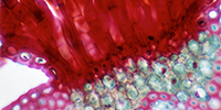
Anatomy is foundational to many branches of science, but students often find morphology to be one of the most difficult subjects they are required to master to become excellent physicians, dentists, physical therapists, nurses, functional morphologists, veterinarians, paleontologists, etc. While dissection labs are invaluable, they are problematic due to expense, exposure to preservatives, and ethical issues of using animals for specimens. Students often struggle with conventional lectures that reduce three-dimensional, complex structures to two-dimensional figures. The aim of this project is to create novel, deeply engaging learning opportunities by developing virtual reality (VR) tools that supplement or replace dissection labs. We developed methods that allow students to use VR to interact with radiological data in ways that enhance the learning of three-dimensional structures, and that take advantage of VR multiplayer capacities — the ability of students and instructors to interact in a 'virtual' space while being physically separated. Serendipitously, our project was a lifesaver during the pandemic, enabling students to continue to interact with each other and the instructors in a virtual classroom. We used these tools and newly developed curricula for the first time in the class BIOLOGY 3665: Form, Function, and Adaptation in Animals with highly positive student responses.
Collaborators
Colleen Farmer
College of Science
School of Biological Sciences
Project Owner
Mark Durham
School of Dentistry
School of Dentistry
Kathryn Moore
School of Medicine
Neurobiology & Anatomy
Project Info
Funded Project Amount$30K
Keywords
Virtual reality in medicine, augmented reality, student engagement, active learning, distance learning, health sciences education, anatomical sciences, functional morphology
Project Status
Funded 2020
Poster
View poster (pdf)